

Antique Anatomical Skull | Genuine Human Specimen on Wood Stand | Circa 19th Century
Couldn't load pickup availability


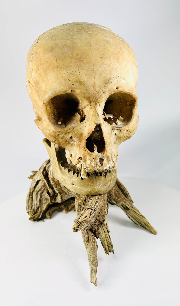
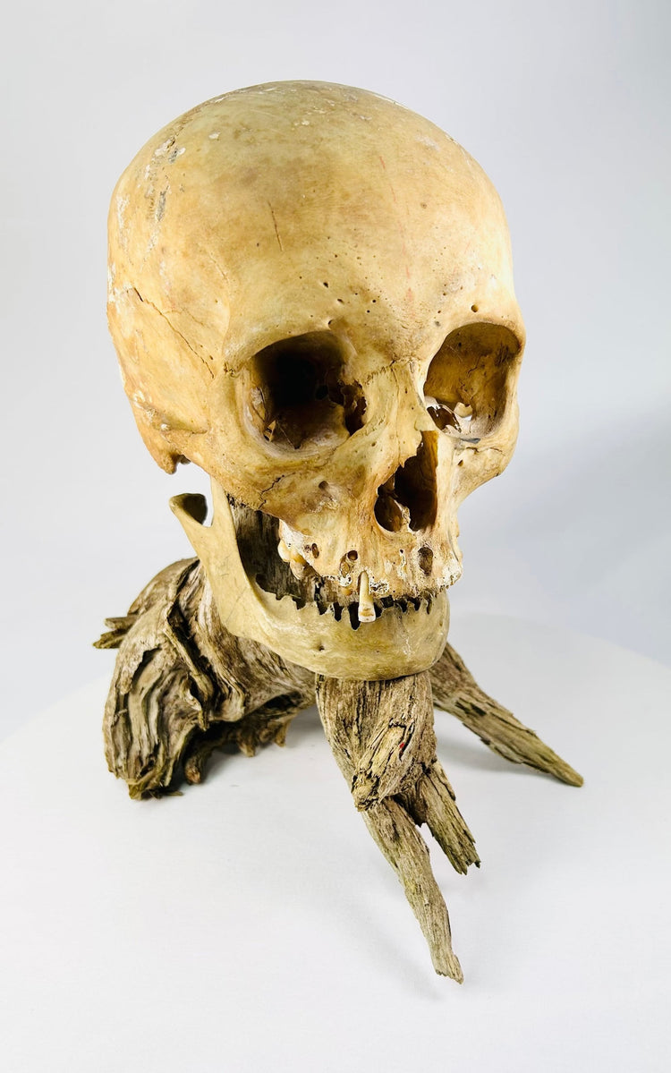
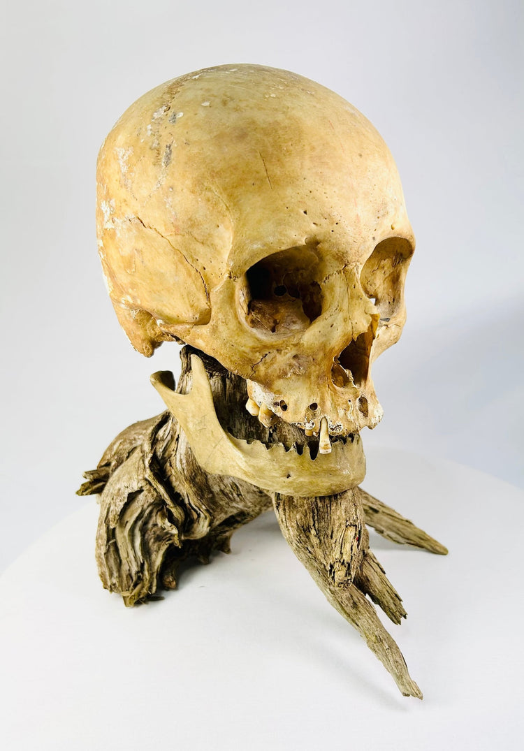
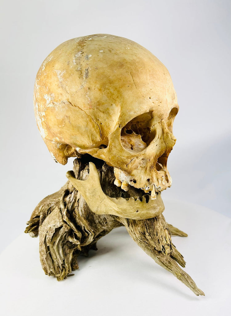
Antique Anatomical Skull | Genuine Human Specimen on Wood Stand | Circa 19th Century
Description
More
Less
Historical Context & Origin
Region: Europe or North America
Material: Human bone, natural driftwood
Period: Circa 19th–early 20th century
Description
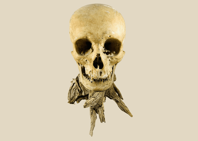
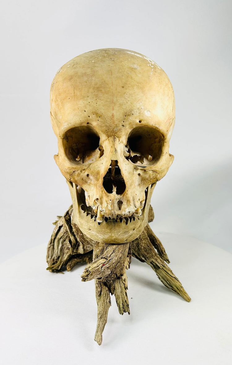
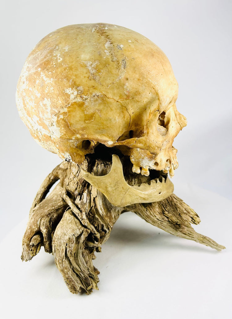
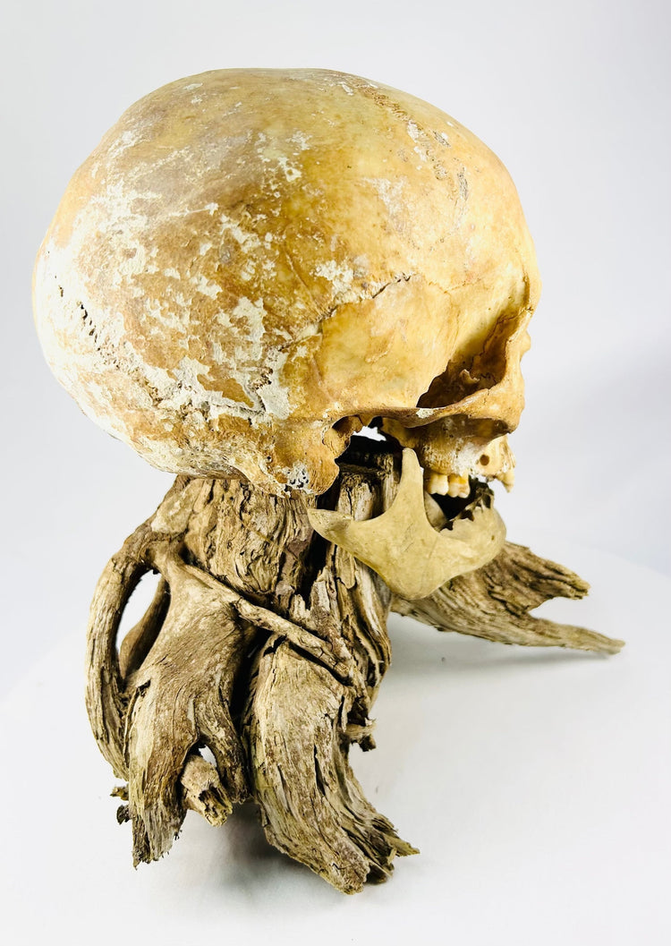
A rare and authentic antique human skull with an associated lower mandible, mounted on an organic driftwood base for stability and study display. This macabre yet scientifically significant specimen reflects the enduring fascination with anatomy and mortality during the 19th and early 20th centuries—a period when medical specimens, anatomical teaching collections, phrenological studies, and cabinets of curiosity were especially prominent.
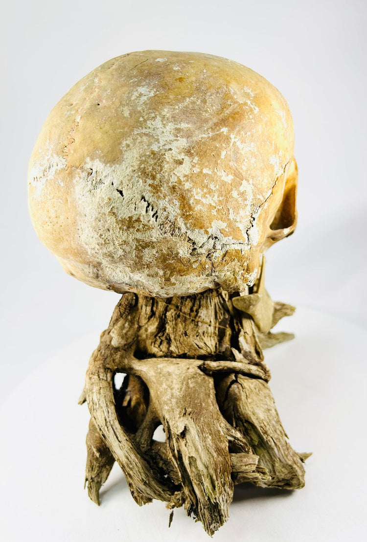
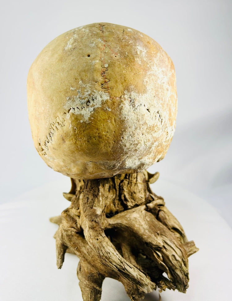
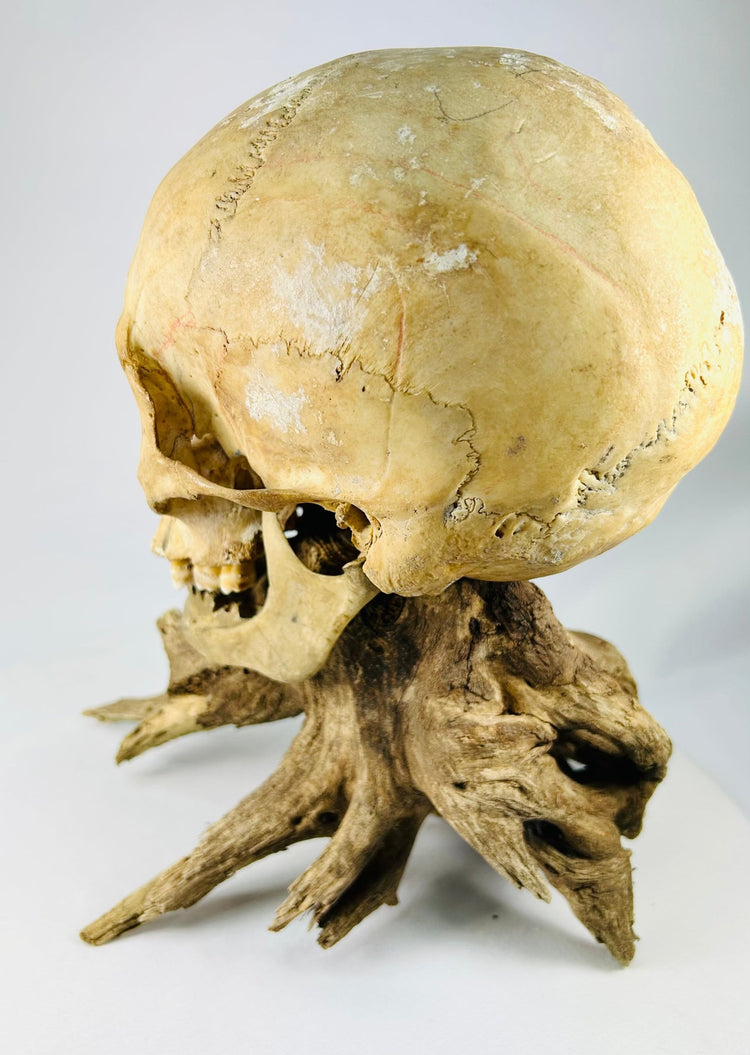
Identified the individual as most likely an adult male. Traits supporting this assessment include the continuation of the zygomatic arch beyond the auricular opening, a pronounced inion at the back of the skull, large and conical mastoid processes, and a slightly sloping frontal bone with projection through the glabellar region between the brow ridges. The lower jaw is comparatively squared, with a pronounced mental eminence, or chin, and flared gonial angles toward the rear.
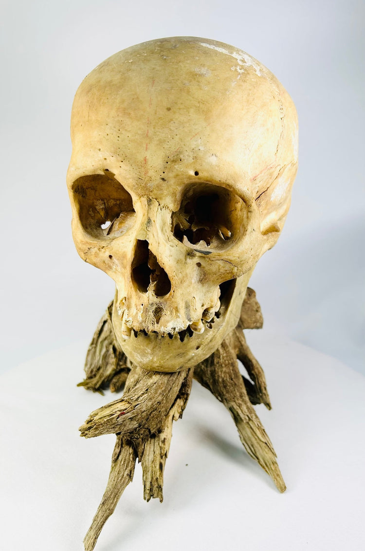
The cranial characteristics were assessed as most consistent with probable European or Caucasian ancestry. The nasal aperture is long and largely straight, with a subtle pear-shaped outline, while the nasal bones appear strongly tented. The nasal spine at the base was likely elongated. Additional traits include rhomboid-shaped and sloping eye orbits, a high cranial vault, and receding malar, or cheek, bones.
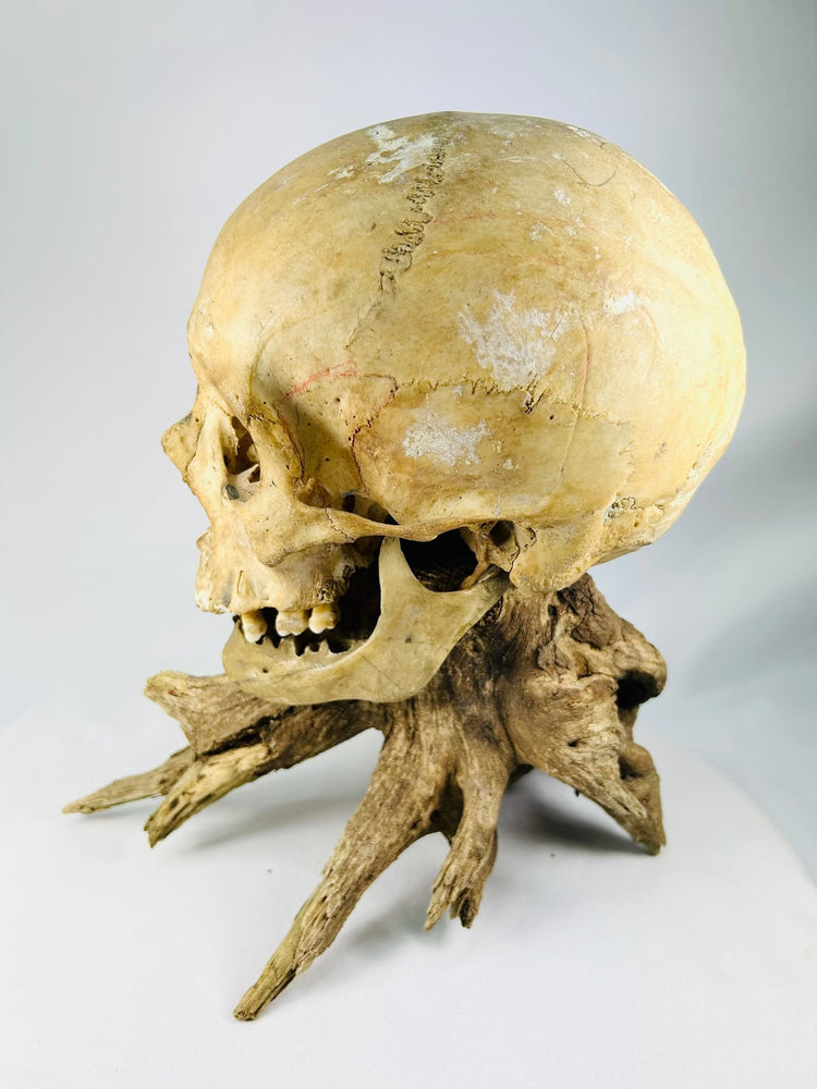
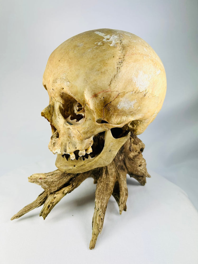
The individual was determined to have been an adult and likely older than 35 years at the time of death. The surviving teeth display considerable wear and deposits of tartar or calculus. Multiple dental abscesses are present, including a substantial abscess in the anterior maxillary, or upper-jaw, region. Several teeth were lost during the individual’s lifetime, as demonstrated by bone resorption in the maxilla, while others were lost after death.
The lateral portions of the coronal suture show fusion associated with the aging process. Although cranial-suture closure is not considered a precise method of determining age on its own, this observation supports the age estimate when considered alongside the extensive dental wear, periodontal disease, abscesses, and ante-mortem tooth loss.
Differences in coloration across portions of the skull were assessed as most likely resulting from post-mortem damage to the outer cortical layer. All of the lower teeth are absent and were likely lost after death, while several missing upper teeth appear to have been lost during life.
The underside of the skull, rear joints, dental alignment, and fit of the jaw were studied to determine whether the mandible and cranium originated from the same person. Their alignment is compatible, allowing for the cartilage that would originally have occupied the joints but does not survive. The corresponding arch shapes and the presence of substantial periodontal disease in both the upper and lower jaws further support the conclusion that the cranium and mandible most likely belonged to the same individual.
Mounted on naturally weathered driftwood, the specimen evokes the anatomical teaching collections, medical displays, and private cabinets of curiosity that became especially prominent during the 19th and early 20th centuries.
Features
- Antique human cranium with associated lower mandible
- Most likely an adult male of probable European or Caucasian ancestry
- Likely older than 35 years at the time of death
- Pronounced inion and large, conical mastoid processes
- Slightly sloping frontal bone with a projected glabellar region
- Squared mandible with a pronounced chin and flared gonial angles
- Long, largely straight nasal aperture with a subtle pear-shaped outline
- Tented nasal bones and a likely elongated nasal spine
- Rhomboid-shaped, sloping eye orbits
- High cranial vault and receding cheekbones
- Significant dental wear and calculus deposits
- Multiple dental abscesses, including a large anterior maxillary abscess
- Evidence of periodontal disease and ante-mortem tooth loss
- Additional teeth lost following death
- Coronal-suture fusion consistent with the aging process
- Cranium and mandible assessed as most likely belonging to the same individual
- Mounted on a naturally weathered driftwood base for stability and presentation
Cultural Significance
During the 19th and early 20th centuries, human skeletal remains were frequently retained by medical universities, teaching hospitals, anatomical museums, and private scientific collections. They served as essential resources for studying anatomy, pathology, human variation, and the physical effects of disease and aging.
The condition of this individual’s dentition provides particularly tangible evidence of historical oral health. The extensive tooth wear, calculus, periodontal disease, abscesses, and tooth loss preserve a physical record of the medical challenges experienced during life.
Today, responsibly documented historical anatomical specimens offer insight into the development of medical education and the changing ways in which the human body has been studied, collected, and interpreted. This example occupies a compelling space between anatomical study, medical history, and the tradition of the cabinet of curiosity.
Condition
Well preserved overall, with expected age-related wear, natural fissures, incomplete dentition, areas of surface alteration, and patination consistent with age.
The surviving teeth display substantial wear and calculus. Multiple dental abscesses are present, including a prominent abscess in the anterior upper jaw. Evidence of periodontal disease, bone resorption, and teeth lost during the individual’s lifetime is visible in the maxilla. Other teeth were lost after death, including all of the lower dentition.
Areas of differing coloration are likely associated with post-mortem damage to the skull’s outer cortical layer. The fit of the mandible is not perfectly flush because the original cartilage of the joints has not survived; however, the alignment, arch shapes, and corresponding periodontal disease support the conclusion that the jaw and cranium most likely belonged to the same person.
The driftwood base displays stable natural weathering and remains structurally sound. Please review all photographs carefully, as they form an important part of the condition description.
Dimensions (Approximate)
Height: 10 in
Width: 6 in
Age (Approximate)
100–150 years old
Provenance
Formerly part of a private collection in Selborne, Hampshire, United Kingdom, where the specimen was preserved among historical, anatomical, and natural-history material.
It subsequently entered a private collection in Toronto, Ontario, Canada, whose interests included antique medical specimens, scientific curiosities, and objects associated with the history of anatomical study.
The specimen was later independently studied by Elizabeth Weiss, PhD, Professor Emerita of Anthropology. Dr. Weiss is a physical anthropologist and bioarchaeologist specializing in human osteology, skeletal remains, paleopathology, and the study of health, disease, trauma, and biological variation through the human skeleton.
Learn More
Read More About Human Medical Specimens: The Fascinating History of Medical Skeletons
View Other Medical Specimens: Skeletal Hand and Skeletal Foot
Description
Historical Context & Origin
Region: Europe or North America
Material: Human bone, natural driftwood
Period: Circa 19th–early 20th century
Description
A rare and authentic antique human skull with an associated lower mandible, mounted on an organic driftwood base for stability and study display. This macabre yet scientifically significant specimen reflects the enduring fascination with anatomy and mortality during the 19th and early 20th centuries—a period when medical specimens, anatomical teaching collections, phrenological studies, and cabinets of curiosity were especially prominent.
Identified the individual as most likely an adult male. Traits supporting this assessment include the continuation of the zygomatic arch beyond the auricular opening, a pronounced inion at the back of the skull, large and conical mastoid processes, and a slightly sloping frontal bone with projection through the glabellar region between the brow ridges. The lower jaw is comparatively squared, with a pronounced mental eminence, or chin, and flared gonial angles toward the rear.
The cranial characteristics were assessed as most consistent with probable European or Caucasian ancestry. The nasal aperture is long and largely straight, with a subtle pear-shaped outline, while the nasal bones appear strongly tented. The nasal spine at the base was likely elongated. Additional traits include rhomboid-shaped and sloping eye orbits, a high cranial vault, and receding malar, or cheek, bones.
The individual was determined to have been an adult and likely older than 35 years at the time of death. The surviving teeth display considerable wear and deposits of tartar or calculus. Multiple dental abscesses are present, including a substantial abscess in the anterior maxillary, or upper-jaw, region. Several teeth were lost during the individual’s lifetime, as demonstrated by bone resorption in the maxilla, while others were lost after death.
The lateral portions of the coronal suture show fusion associated with the aging process. Although cranial-suture closure is not considered a precise method of determining age on its own, this observation supports the age estimate when considered alongside the extensive dental wear, periodontal disease, abscesses, and ante-mortem tooth loss.
Differences in coloration across portions of the skull were assessed as most likely resulting from post-mortem damage to the outer cortical layer. All of the lower teeth are absent and were likely lost after death, while several missing upper teeth appear to have been lost during life.
The underside of the skull, rear joints, dental alignment, and fit of the jaw were studied to determine whether the mandible and cranium originated from the same person. Their alignment is compatible, allowing for the cartilage that would originally have occupied the joints but does not survive. The corresponding arch shapes and the presence of substantial periodontal disease in both the upper and lower jaws further support the conclusion that the cranium and mandible most likely belonged to the same individual.
Mounted on naturally weathered driftwood, the specimen evokes the anatomical teaching collections, medical displays, and private cabinets of curiosity that became especially prominent during the 19th and early 20th centuries.
Features
- Antique human cranium with associated lower mandible
- Most likely an adult male of probable European or Caucasian ancestry
- Likely older than 35 years at the time of death
- Pronounced inion and large, conical mastoid processes
- Slightly sloping frontal bone with a projected glabellar region
- Squared mandible with a pronounced chin and flared gonial angles
- Long, largely straight nasal aperture with a subtle pear-shaped outline
- Tented nasal bones and a likely elongated nasal spine
- Rhomboid-shaped, sloping eye orbits
- High cranial vault and receding cheekbones
- Significant dental wear and calculus deposits
- Multiple dental abscesses, including a large anterior maxillary abscess
- Evidence of periodontal disease and ante-mortem tooth loss
- Additional teeth lost following death
- Coronal-suture fusion consistent with the aging process
- Cranium and mandible assessed as most likely belonging to the same individual
- Mounted on a naturally weathered driftwood base for stability and presentation
Cultural Significance
During the 19th and early 20th centuries, human skeletal remains were frequently retained by medical universities, teaching hospitals, anatomical museums, and private scientific collections. They served as essential resources for studying anatomy, pathology, human variation, and the physical effects of disease and aging.
The condition of this individual’s dentition provides particularly tangible evidence of historical oral health. The extensive tooth wear, calculus, periodontal disease, abscesses, and tooth loss preserve a physical record of the medical challenges experienced during life.
Today, responsibly documented historical anatomical specimens offer insight into the development of medical education and the changing ways in which the human body has been studied, collected, and interpreted. This example occupies a compelling space between anatomical study, medical history, and the tradition of the cabinet of curiosity.
Condition
Well preserved overall, with expected age-related wear, natural fissures, incomplete dentition, areas of surface alteration, and patination consistent with age.
The surviving teeth display substantial wear and calculus. Multiple dental abscesses are present, including a prominent abscess in the anterior upper jaw. Evidence of periodontal disease, bone resorption, and teeth lost during the individual’s lifetime is visible in the maxilla. Other teeth were lost after death, including all of the lower dentition.
Areas of differing coloration are likely associated with post-mortem damage to the skull’s outer cortical layer. The fit of the mandible is not perfectly flush because the original cartilage of the joints has not survived; however, the alignment, arch shapes, and corresponding periodontal disease support the conclusion that the jaw and cranium most likely belonged to the same person.
The driftwood base displays stable natural weathering and remains structurally sound. Please review all photographs carefully, as they form an important part of the condition description.
Dimensions (Approximate)
Height: 10 in
Width: 6 in
Age (Approximate)
100–150 years old
Provenance
Formerly part of a private collection in Selborne, Hampshire, United Kingdom, where the specimen was preserved among historical, anatomical, and natural-history material.
It subsequently entered a private collection in Toronto, Ontario, Canada, whose interests included antique medical specimens, scientific curiosities, and objects associated with the history of anatomical study.
The specimen was later independently studied by Elizabeth Weiss, PhD, Professor Emerita of Anthropology. Dr. Weiss is a physical anthropologist and bioarchaeologist specializing in human osteology, skeletal remains, paleopathology, and the study of health, disease, trauma, and biological variation through the human skeleton.
Learn More
Read More About Human Medical Specimens: The Fascinating History of Medical Skeletons
View Other Medical Specimens: Skeletal Hand and Skeletal Foot
You May Also Like
